Page Not Found
Page not found. Your pixels are in another canvas.
A list of all the posts and pages found on the site. For you robots out there is an XML version available for digesting as well.
Page not found. Your pixels are in another canvas.
This is a page not in th emain menu
Published:
This post will show up by default. To disable scheduling of future posts, edit config.yml and set future: false.
Published:
This is a sample blog post. Lorem ipsum I can’t remember the rest of lorem ipsum and don’t have an internet connection right now. Testing testing testing this blog post. Blog posts are cool.
Published:
This is a sample blog post. Lorem ipsum I can’t remember the rest of lorem ipsum and don’t have an internet connection right now. Testing testing testing this blog post. Blog posts are cool.
Published:
This is a sample blog post. Lorem ipsum I can’t remember the rest of lorem ipsum and don’t have an internet connection right now. Testing testing testing this blog post. Blog posts are cool.
Published:
This is a sample blog post. Lorem ipsum I can’t remember the rest of lorem ipsum and don’t have an internet connection right now. Testing testing testing this blog post. Blog posts are cool.
Published in Cell, 2016
Branched actin networks—created by the Arp2/3 complex, capping protein, and a nucleation promoting factor—generate and transmit forces required for many cellular processes, but their response to force is poorly understood. To address this, we assembled branched actin networks in vitro from purified components and used simultaneous fluorescence and atomic force microscopy to quantify their molecular composition and material properties under various forces.
Download here
Published in Optics Express, 2016
Using light-sheet microscopy combined with digital Fourier methods we probe the dynamics of colloidal samples and DNA molecules. This combination, referred to as selective-plane illumination differential dynamic microscopy (SPIDDM), has the benefit of optical sectioning to study, with minimal photobleaching, thick samples allowing us to measure the diffusivity of colloidal particles at high volume fractions.
Download here
Published in Soft Matter, 2016
Previous experiments have shown that spherical colloidal particles relax to equilibrium slowly after they adsorb to a liquid–liquid interface, despite the large interfacial energy gradient driving the adsorption. The slow relaxation has been explained in terms of transient pinning and depinning of the contact line on the surface of the particles. However, the nature of the pinning sites has not been investigated in detail. We use digital holographic microscopy to track a variety of colloidal spheres—inorganic and organic, charge-stabilized and sterically stabilized, aqueous and non-aqueous—as they breach liquid interfaces.
Download here
Published in Optics Express, 2017
Selective-plane illumination microscopy (SPIM) provides unparalleled advantages for the volumetric imaging of living organisms over extended times. However, the spatial configuration of a SPIM system often limits its compatibility with many widely used biological sample holders such as multi-well chambers and plates. To solve this problem, we developed a high numerical aperture (NA) open-top configuration that places both the excitation and detection objectives on the opposite of the sample coverglass.
Download here
Published in Nature Cell Biology, 2017
Ciliopathies, including nephronophthisis (NPHP), Meckel syndrome (MKS) and Joubert syndrome (JBTS), can be caused by mutations affecting components of the transition zone, a domain near the base of the cilium that controls the protein composition of its membrane. We defined the three-dimensional arrangement of key proteins in the transition zone using two-colour stochastic optical reconstruction microscopy (STORM).
Download here
Published in Optics Letters, 2017
While numerous optical methods exist to probe the dynamics of biological or complex fluid samples, in recent years digital Fourier microscopy techniques such as differential dynamic microscopy have emerged as ways to efficiently combine elements of imaging and scattering methods. Here, we demonstrate, through experiments and simulations, how point-spread function engineering can be used to extend the reach of differential dynamic microscopy.
Download here
Published in Soft Matter, 2019
Crowding plays a key role in the transport and conformations of biological macromolecules. Gene therapy, viral infection, and transfection require DNA to traverse the crowded cytoplasm, including the cytoskeletal network of filamentous proteins. Given the complexity of cellular crowding, the dynamics of biological molecules can be highly dependent on the spatiotemporal scale probed. We present a powerful platform that spans molecular and cellular scales by coupling single-molecule conformational tracking (SMCT) and selective-plane illumination differential dynamic microscopy (SPIDDM).
Download here
Published in Soft Matter, 2019
The interface between two fluids is roughened by thermally excited capillary waves. By using colloid-polymer systems which exhibit liquid-gas phase separation, the time and length scales of capillary waves become accessible to optical microscopy methods. Here, we study such a system using bright-field optical microscopy combined with a novel extension of differential dynamic microscopy. =
Download here
Published in Physical Review E, 2019
Particles bound to fluid-fluid interfaces are widely used to study self-assembly and to make materials such as Pickering emulsions. In both contexts, the lateral interactions between such particles have been studied extensively. However, much less is known about the normal interactions between a particle and the interface prior to contact. We use digital holographic microscopy to measure the dynamics of individual micrometer-size colloidal particles as they approach an interface between an aqueous phase and oil.
Download here
Published in Biomacromolecules, 2019
The diffusion of microscopic particles through the cell, important to processes such as viral infection, gene delivery, and vesicle transport, is largely controlled by the complex cytoskeletal network, comprised of semiflexible actin filaments and rigid microtubules, that pervades the cytoplasm. By varying the relative concentrations of actin and microtubules, the cytoskeleton can display a host of different structural and dynamic properties that, in turn, impact the diffusion of particles through the composite network. Here, we couple single-particle tracking with differential dynamic microscopy to characterize the transport of microsphere tracers diffusing through composite in vitro networks with varying ratios of actin and microtubules.
Download here
Published in Science Advances, 2019
Cytoskeletal crowding plays a key role in the diffusion of DNA molecules through the cell, acting as a barrier to effective intracellular transport and conformational stability required for processes such as transfection, viral infection, and gene therapy. Here, we elucidate the transport properties and conformational dynamics of linear and ring DNA molecules diffusing through entangled and crosslinked composite networks of actin and microtubules.
Download here
Published in The Biophysicist, 2020
Recent work has shown that the intracellular environment is organized not only through membrane-bound organelles but also through fluid droplets that emerge through liquid-liquid phase separation (LLPS). Intracellular LLPS has attracted recent attention because these fluid droplets, termed biomolecular condensates or membraneless organelles, seem to play important roles in cells’ responses to stress, gene regulation, and pathologies. Our understanding of intracellular LLPS has advanced through many quantitative biophysical techniques. Here, we describe a set of undergraduate lab activities that highlight these biophysical techniques.
Download here
Published in Soft Matter, 2020
The cytoskeleton, a complex network of protein filaments and crosslinking proteins, dictates diverse cellular processes ranging from division to cargo transport. Yet, the role the cytoskeleton plays in the intracellular transport of DNA and other macromolecules remains poorly understood. Here, using single-molecule conformational tracking, we measure the transport and conformational dynamics of linear and relaxed circular (ring) DNA in composite networks of actin and microtubules with variable types of crosslinking.
Download here
Published in Review of Scientific Instruments, 2021
Differential dynamic microscopy (DDM) is increasingly used in the fields of soft matter physics and biophysics to extract the dynamics of microscopic objects across a range of wavevectors by optical microscopy. Standard DDM is limited to detecting dynamics no faster than the camera frame rate. We report on an extension to DDM where we sequentially illuminate the sample with spectrally distinct light and image with a color camera. By pulsing blue and then red light separated by a lag time much smaller than the camera’s exposure time, we are able to use this two-color DDM method to measure dynamics occurring much faster than the camera frame rate.
Recommended citation: R. You and R. McGorty (2021). "Two-color differential dynamic microscopy for capturing fast dynamics" Review of Scientific Instruments. 1(1). https://aip.scitation.org/doi/abs/10.1063/5.0039177
Published in Science Advances, 2021
The cytoskeleton is a dynamic network of proteins, including actin, microtubules, and their associated motor proteins, that enables essential cellular processes such as motility, division, and growth. While actomyosin networks are extensively studied, how interactions between actin and microtubules, ubiquitous in the cytoskeleton, influence actomyosin activity remains an open question. Here, we create a network of co-entangled actin and microtubules driven by myosin II. We combine dynamic differential microscopy, particle image velocimetry, and particle tracking to show that both actin and microtubules undergo ballistic contraction with unexpectedly indistinguishable characteristics.
Download here
Published in Soft Matter, 2021
Anomalous diffusion in crowded and complex environments is widely studied due to its importance in intracellular transport, fluid rheology and materials engineering. Specifically, diffusion through the cytoskeleton, a network comprised of semiflexible actin filaments and rigid microtubules that interact both sterically and via crosslinking, plays a principal role in viral infection, vesicle transport and targeted drug delivery. Here, we elucidate the impact of crosslinking on particle diffusion in composites of actin and microtubules with actin-actin, microtubule-microtubule and actin-microtubule crosslinking.
Download here
Published in Soft Matter, 2021
Microcapsules allow for the controlled containment, transport, and release of cargoes ranging from pharmaceuticals to fragrances. Given the interest from a variety of industries in microcapsules and other core-shell structures, a multitude of fabrication strategies exist. Here, we report on a method relying on a mixture of temperature-responsive microgel particles, poly(N-isopropylacrylamide) (pNIPAM), and a polymer which undergo fluid-fluid phase separation.
Download here
Published in Frontiers in Physics, 2021
Volumetric microscopic imaging data acquired at high speeds is often needed in studies of soft matter. Several microscopy techniques exist for this purpose, but a relative newcomer is light sheet fluorescence microscopy (LSFM). This microscopy method has seen spectacular growth in the biological sciences over the past two decades. In this perspective, we highlight how LSFM may also apply to the field of soft matter. We review the principles and recent advances of LSFM and discuss how it has been used in prior soft matter studies. We demonstrate how a recent implementation of LSFM can be used to study capillary wave fluctuations and droplet coalescence in a colloidal fluid system.
Download here
Published in Soft Matter, 2021
The cytoskeleton is a model active matter system that controls processes as diverse as cell motility and mechanosensing. While both active actomyosin dynamics and actin–microtubule interactions are key to the cytoskeleton’s versatility and adaptability, an understanding of their interplay is lacking. Here, we couple microscale experiments with mechanistic modeling to elucidate how connectivity, rigidity, and force-generation affect emergent material properties in composite networks of actin, tubulin, and myosin. We use multi-spectral imaging, time-resolved differential dynamic microscopy and spatial image autocorrelation to show that ballistic contraction occurs in composites with sufficient flexibility and motor density, but that a critical fraction of microtubules is necessary to sustain controlled dynamics.
Download here
Published in Journal of Rheology, 2022
Polymer architecture plays critical roles in both bulk rheological properties and microscale macromolecular dynamics in entangled polymer solutions and composites. Ring polymers, in particular, have been the topic of much debate due to the inability of the celebrated reptation model to capture their observed dynamics. Macrorheology and differential dynamic microscopy (DDM) are powerful methods to determine entangled polymer dynamics across scales; yet, they typically require different samples under different conditions, preventing direct coupling of bulk rheological properties to the underlying macromolecular dynamics. Here, we perform macrorheology on composites of highly overlapping DNA and dextran polymers, focusing on the role of DNA topology (rings versus linear chains) as well as the relative volume fractions of DNA and dextran. On the same samples under the same conditions, we perform DDM and single-molecule tracking on embedded fluorescent-labeled DNA molecules immediately before and after bulk measurements. We show DNA-dextran composites exhibit unexpected nonmonotonic dependences of bulk viscoelasticity and molecular-level transport properties on the fraction of DNA comprising the composites, with characteristics that are strongly dependent on the DNA topology. We rationalize our results as arising from stretching and bundling of linear DNA versus compaction, swelling, and threading of rings driven by dextran-mediated depletion interactions.
Download here
Published in JoVE, 2022
Cells can crawl, self-heal, and tune their stiffness due to their remarkably dynamic cytoskeleton. As such, reconstituting networks of cytoskeletal biopolymers may lead to a host of active and adaptable materials. However, engineering such materials with precisely tuned properties requires measuring how the dynamics depend on the network composition and synthesis methods. Quantifying such dynamics is challenged by variations across the time, space, and formulation space of composite networks. The protocol here describes how the Fourier analysis technique, differential dynamic microscopy (DDM), can quantify the dynamics of biopolymer networks and is particularly well suited for studies of cytoskeleton networks. DDM works on time sequences of images acquired using a range of microscopy modalities, including laser-scanning confocal, widefield fluorescence, and brightfield imaging. From such image sequences, one can extract characteristic decorrelation times of density fluctuations across a span of wave vectors. A user-friendly, open-source Python package to perform DDM analysis is also developed. With this package, one can measure the dynamics of labeled cytoskeleton components or of embedded tracer particles, as demonstrated here with data of intermediate filament (vimentin) networks and active actin-microtubule networks. Users with no prior programming or image processing experience will be able to perform DDM using this software package and associated documentation.
Download here
Published in Frontiers in Physics, 2022
Shear driven patterning is seen in many soft matter systems. We use rheology and optical microscopy to probe the structures formed when we shear a colloid-polymer mixture containing temperature-sensitive microgel particles. By increasing the temperature, we can increase the particle attraction and transition from liquid-like to gel-like behavior. And by applying shear flow to the sample as the temperature and, hence, state of the system changes, we can affect the morphology of mesoscopic colloidal clusters. We can produce gels comprised of fibrous, elongated colloid-rich clusters, or we can form more isotropic clusters. The rheology is measured and shear-induced flocculation observed for colloid-polymer systems with different cluster morphologies. At shear rates high enough to produce elongated clusters but low enough to not break clusters apart, we observe log-like flocs that are aligned with the vorticity direction and roll between the parallel plates of our rheometer.
Download here
Published in JoVE, 2022
The composite cytoskeleton, comprising interacting networks of semiflexible actin filaments and rigid microtubules, restructures and generates forces using motor proteins such as myosin II and kinesin to drive key processes such as migration, cytokinesis, adhesion, and mechanosensing. While actin-microtubule interactions are key to the cytoskeleton’s versatility and adaptability, an understanding of their interplay with myosin and kinesin activity is still nascent. This work describes how to engineer tunable three-dimensional composite networks of co-entangled actin filaments and microtubules that undergo active restructuring and ballistic motion, driven by myosin II and kinesin motors, and are tuned by the relative concentrations of actin, microtubules, motor proteins, and passive crosslinkers. Protocols for fluorescence labeling of the microtubules and actin filaments to most effectively visualize composite restructuring and motion using multi-spectral confocal imaging are also detailed. Finally, the results of data analysis methods that can be used to quantitatively characterize non-equilibrium structure, dynamics, and mechanics are presented. Recreating and investigating this tunable biomimetic platform provides valuable insight into how coupled motor activity, composite mechanics, and filament dynamics can lead to myriad cellular processes from mitosis to polarization to mechano-sensation.
Download here
Published in Nature Communications, 2022
How local stresses propagate through polymeric fluids, and, more generally, how macromolecular dynamics give rise to viscoelasticity are open questions vital to wide-ranging scientific and industrial fields. Here, to unambiguously connect polymer dynamics to force response, and map the deformation fields that arise in macromolecular materials, we present Optical-Tweezers-integrating-Differential -Dynamic-Microscopy (OpTiDMM) that simultaneously imposes local strains, measures resistive forces, and analyzes the motion of the surrounding polymers. Our measurements with blends of ring and linear polymers (DNA) and their composites with stiff polymers (microtubules) uncover an unexpected resonant response, in which strain alignment, superdiffusivity, and elasticity are maximized when the strain rate is comparable to the entanglement rate. Microtubules suppress this resonance, while substantially increasing elastic storage, due to varying degrees to which the polymers buildup, stretch and flow along the strain path, and configurationally relax induced stress. More broadly, the rich multi-scale coupling of mechanics and dynamics afforded by OpTiDDM, empowers its interdisciplinary use to elucidate non-trivial phenomena that sculpt stress propagation dynamics–critical to commercial applications and cell mechanics alike.
Download here
Published in iScience, 2022
Dynamics of biological macromolecules, such as DNA, in crowded and confined environments are critical to understanding cellular processes such as transcription, infection, and replication. However, the combined effects of cellular confinement and crowding on macromolecular dynamics remain poorly understood. Here, we use differential dynamic microscopy to investigate the diffusion of large DNA molecules confined in cell-sized droplets and crowded by dextran polymers. We show that confined and crowded DNA molecules exhibit universal anomalous subdiffusion with scaling that is insensitive to the degree of confinement and crowding. However, effective DNA diffusion coefficients Deff decrease up to 2 orders of magnitude as droplet size decreases–an effect that is enhanced by increased crowding. We mathematically model the coupling of crowding and confinement by combining polymer scaling theories with confinement-induced depletion effects. The generality and tunability of our system and models render them applicable to elucidating wide-ranging crowded and confined systems.
Download here
Published in Frontiers in Physics, 2022
The cytoskeleton–a composite network of biopolymers, molecular motors, and associated binding proteins–is a paradigmatic example of active matter. Particle transport through the cytoskeleton can range from anomalous and heterogeneous subdiffusion to superdiffusion and advection. Yet, recapitulating and understanding these properties–ubiquitous to the cytoskeleton and other out-of-equilibrium soft matter systems–remains challenging. Here, we combine light sheet microscopy with differential dynamic microscopy and single-particle tracking to elucidate anomalous and advective transport in actomyosin-microtubule composites. We show that particles exhibit multi-mode transport that transitions from pronounced subdiffusion to superdiffusion at tunable crossover timescales. Surprisingly, while higher actomyosin content increases the range of timescales over which transport is superdiffusive, it also markedly increases the degree of subdiffusion at short timescales and generally slows transport. Corresponding displacement distributions display unique combinations of non-Gaussianity, asymmetry, and non-zero modes, indicative of directed advection coupled with caged diffusion and hopping. At larger spatiotemporal scales, particles in active composites exhibit superdiffusive dynamics with scaling exponents that are robust to changing actomyosin fractions, in contrast to normal, yet faster, diffusion in networks without actomyosin. Our specific results shed important new light on the interplay between non-equilibrium processes, crowding and heterogeneity in active cytoskeletal systems. More generally, our approach is broadly applicable to active matter systems to elucidate transport and dynamics across scales.
Download here
Published in Advanced Materials, 2023
Polymer topology, which plays a principal role in the rheology of polymeric fluids, and non-equilibrium materials, which exhibit time-varying rheological properties, are topics of intense investigation. Here, we push composites of circular DNA and dextran out-of-equilibrium via enzymatic digestion of DNA rings to linear fragments. Our time-resolved rheology measurements reveal discrete state-switching, with composites undergoing abrupt transitions between dissipative and elastic-like states. The gating time and lifetime of the elastic-like states, and the magnitude and sharpness of the transitions, are surprisingly decorrelated from digestion rates and non-monotonically depend on the DNA fraction. We model our results using sigmoidal two-state functions to show that bulk state-switching can arise from continuous molecular-level activity due to the necessity for cooperative percolation of entanglements to support macroscopic stresses. Our platform, coupling the tunability of topological composites with the power of enzymatic reactions, may be leveraged for diverse material applications from wound-healing to self-repairing infrastructure.
Download here
Published in PNAS Nexus, 2023
The cellular cytoskeleton relies on diverse populations of motors, filaments, and binding proteins acting in concert to enable non-equilibrium processes ranging from mitosis to chemotaxis. The cytoskeleton’s versatile reconfigurability, programmed by interactions between its constituents, make it a foundational active matter platform. However, current active matter endeavors are limited largely to single force-generating components acting on a single substrate–far from the composite cytoskeleton in cells. Here, we engineer actin-microtubule composites, driven by kinesin and myosin motors and tuned by crosslinkers, to ballistically restructure and flow with speeds that span three orders of magnitude depending on the composite formulation and time relative to the onset of motor activity. Differential dynamic microscopy analyses reveal that kinesin and myosin compete to delay the onset of acceleration and suppress discrete restructuring events, while passive crosslinking of either actin or microtubules has an opposite effect. Our minimal advection-diffusion model and spatial correlation analyses correlate these dynamics to structure, with motor antagonism suppressing reconfiguration and de-mixing, while crosslinking enhances clustering. Despite the rich formulation space and emergent formulation-dependent structures, the non-equilibrium dynamics across all composites and timescales can be organized into three classes–slow isotropic reorientation, fast directional flow, and multimode restructuring. Moreover, our mathematical model demonstrates that diverse structural motifs can arise simply from the interplay between motor-driven advection and frictional drag. These general features of our platform facilitate applicability to other active matter systems, and shed light on diverse ways that cytoskeletal components can cooperate or compete to enable wide-ranging cellular processes.
Download here
Published in JoVE, 2024
Reconstituted cytoskeleton composites have emerged as a valuable model system for studying non-equilibrium soft matter. The faithful capture of the dynamics of these 3D, dense networks calls for optical sectioning, which is often associated with fluorescence confocal microscopes. However, recent developments in light-sheet fluorescence microscopy (LSFM) have established it as a cost-effective and, at times, superior alternative. To make LSFM accessible to cytoskeleton researchers less familiar with optics, we present a step-by-step beginner’s guide to building a versatile light-sheet fluorescence microscope from off-the-shelf components. To enable sample mounting with traditional slide samples, this LSFM follows the single-objective light-sheet (SOLS) design, which utilizes a single objective for both the excitation and emission collection. We describe the function of each component of the SOLS in…
Download here
Published in Soft Matter, 2024
DNA, which naturally occurs in linear, ring, and supercoiled topologies, frequently undergoes enzyme-driven topological conversion and fragmentation in vivo, enabling it to perform a variety of functions within the cell. In vitro, highly concentrated DNA polymers form entanglements that yield viscoelastic properties dependent on the topologies and lengths of the DNA. Enzyme-driven alterations of DNA size and shape therefore offer a means of designing active materials with programmable viscoelastic properties. Here, we incorporate multi-site restriction endonucleases into dense DNA solutions to linearize and fragment circular DNA molecules. We pair optical tweezers microrheology with differential dynamic microscopy and single-molecule tracking to measure the linear and nonlinear viscoelastic response and transport properties of entangled DNA solutions over a wide range of spatiotemporal scales throughout the course of enzymatic digestion. We show that, at short timescales, relative to the relaxation timescales of the polymers, digestion of these ‘topologically-active’ fluids initially causes an increase in elasticity and relaxation times followed by a gradual decrease. Conversely, for long timescales, linear viscoelastic moduli exhibit signatures of increasing elasticity. DNA diffusion, likewise, becomes increasingly slowed, in direct opposition to the short-time behavior. We hypothesize that this scale-dependent rheology arises from the population of small DNA fragments, which increases as digestion proceeds, driving self-association of larger fragments via depletion interactions, giving rise to slow relaxation modes of clusters of entangled chains, interspersed among shorter unentangled fragments. While these slow modes likely dominate at long times, they are presumably frozen out in the short-time limit, which instead probes the faster relaxation modes of the unentangled population.
Download here
Published in Soft Matter, 2024
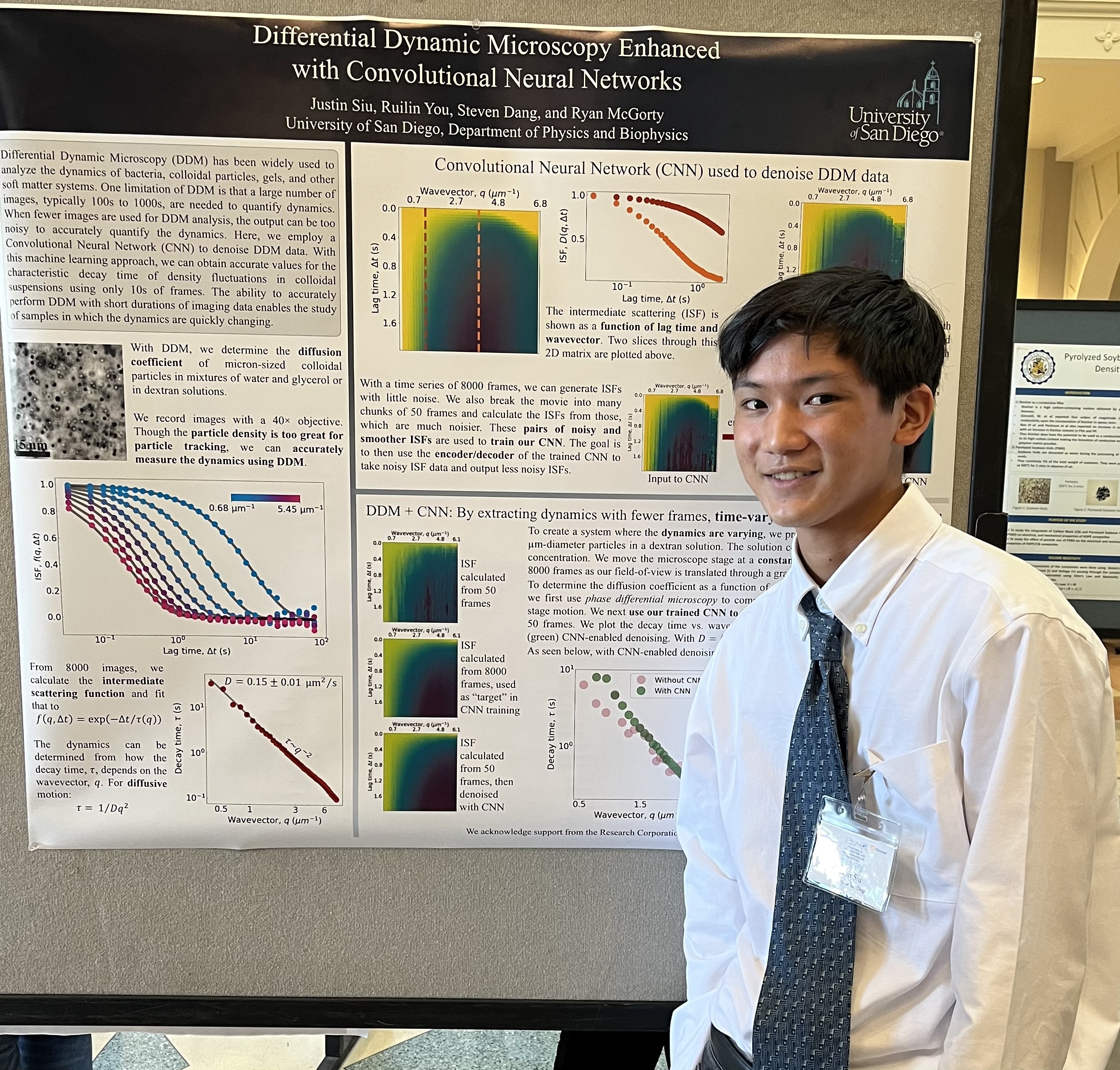
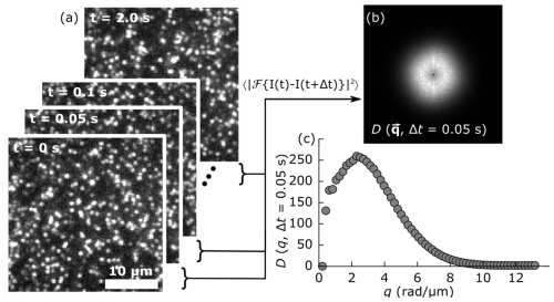
Differential dynamic microscopy (DDM) typically relies on movies containing hundreds or thousands of frames to accurately quantify motion in soft matter systems. Using movies much shorter in duration produces noisier and less accurate results. This limits the applicability of DDM to situations where the dynamics are stationary over extended times. Here, we investigate a method to denoise the DDM process, particularly suited to when a limited number of imaging frames are available or when dynamics are quickly evolving in time. We use a convolutional neural network encoder-decoder (CNN-ED) model to reduce the noise in the intermediate scattering function that is computed via DDM. We demonstrate this approach of combining machine learning and DDM on samples containing diffusing micron-sized colloidal particles. We quantify how the particles’ diffusivities change over time as the fluid they are suspended in gels. We also quantify how the diffusivity of particles varies with position in a sample containing a viscosity gradient. These test cases demonstrate how studies of non-equilibrium dynamics and high-throughput screens could benefit from a method to denoise the outputs of DDM.
Download here
Published in STAR Protocols, 2024
Here, we present a protocol for encapsulating DNA molecules under crowded conditions within cell-sized lipid-coated droplets. We describe steps for preparing a lipid-oil mixture and adding an aqueous solution containing DNA, which, when mixed, forms water-in-oil droplets of radii between ∼5 and 100 μm. We then detail procedures for quantifying the dynamics of DNA molecules in these droplets by analyzing fluorescence microscopy time series using differential dynamic microscopy. This protocol can be utilized to investigate DNA transport within a range of biomimetic and crowded environments.
Download here
Published in Soft Matter, 2024
Blends of circular and linear polymers have fascinated researchers for decades, and the role of topology on their stress response and dynamics remains fervently debated. While linear polymers adopt larger coil sizes and form stronger, more pervasive entanglements than their circular counterparts, threading of circular polymers by linear chains can introduce persistent constraints that dramatically decrease mobility, leading to emergent rheological properties in blends. However, the complex interplay between topology-dependent polymer overlap and threading propensity, along with the large amounts of material required to sample many compositions, has limited the ability to experimentally map stress response to composition with high resolution. Moreover, the role of supercoiling on the response of circular-linear blends remains poorly understood. Here, we leverage in situ enzymatic topological conversion to map the deformation dynamics of DNA blends with over 70 fractions of linear, ring and supercoiled molecules that span the phase space of possible topological compositions. We use OpTiDDM (optical tweezers integrating differential dynamic microscopy) to map strain-induced deformation dynamics to composition, revealing that strain-coupling, quantified by superdiffusive dynamics that are aligned with the strain, is maximized for blends with comparable fractions of ring and linear polymers. Increasing the supercoiled fraction dramatically reduces strain-coupling, while converting rings to linear chains offers more modest coupling reduction. We demonstrate that these results are a direct consequence of the interplay between increasing polymer overlap and decreasing threading probability as circular molecules are converted to linear chains, with a careful balance achieved for blends with ample ring fractions but devoid of supercoiled molecules.
Download here
Published in Acta Biomaterialia, 2024
Blends of circular and linear polymers have fascinated researchers for decades, and the role of topology on their stress response and dynamics remains fervently debated. While linear polymers adopt larger coil sizes and form stronger, more pervasive entanglements than their circular counterparts, threading of circular polymers by linear chains can introduce persistent constraints that dramatically decrease mobility, leading to emergent rheological properties in blends. However, the complex interplay between topology-dependent polymer overlap and threading propensity, along with the large amounts of material required to sample many compositions, has limited the ability to experimentally map stress response to composition with high resolution. Moreover, the role of supercoiling on the response of circular-linear blends remains poorly understood. Here, we leverage in situ enzymatic topological conversion to map the deformation dynamics of DNA blends with over 70 fractions of linear, ring and supercoiled molecules that span the phase space of possible topological compositions. We use OpTiDDM (optical tweezers integrating differential dynamic microscopy) to map strain-induced deformation dynamics to composition, revealing that strain-coupling, quantified by superdiffusive dynamics that are aligned with the strain, is maximized for blends with comparable fractions of ring and linear polymers. Increasing the supercoiled fraction dramatically reduces strain-coupling, while converting rings to linear chains offers more modest coupling reduction. We demonstrate that these results are a direct consequence of the interplay between increasing polymer overlap and decreasing threading probability as circular molecules are converted to linear chains, with a careful balance achieved for blends with ample ring fractions but devoid of supercoiled molecules.
Download here
Published in PRX Life, 2025
The cytoskeleton is an active and complex composite network of actin, microtubules, and intermediate filaments (IFs). Networks of these distinct biopolymers work in partnership to fulfill fundamental cellular processes, such as cell polarization, intracellular transport, and cell migration. While there is evidence that the different cytoskeletal filaments interact through biochemical signaling and physical interactions, many basic questions remain regarding how this crosstalk impacts the dynamics and organization of the cytoskeletal filaments. Here, we combine Fourier image analysis methods with live cell experiments to characterize the impacts of cytoskeletal crosstalk on the structure and dynamics of microtubules in mouse embryonic fibroblasts.
Download here
Published in Nature Communications, 2025
Active, responsive, non-equilibrium materials-at the forefront of materials engineering-offer dynamical restructuring, mobility and other complex life-like properties. Yet, this enhanced functionality comes with significant amplification of the size and complexity of the datasets needed to characterize their properties, thereby challenging conventional approaches to analysis. To meet this need, we present BARCODE: Biomaterial Activity Readouts to Categorize, Optimize, Design and Engineer, an open-access software that automates high throughput screening of microscopy video data to enable non-equilibrium material optimization and discovery. BARCODE produces a unique fingerprint or ‘barcode’ of performance metrics that visually and quantitatively encodes dynamic material properties with minimal file size.
Download here
Published:
 | Convolutional neural networks applied to differential dynamic microscopy reduces noise when quantifying heterogeneous dynamics *Gildardo Martinez*, *Justin Siu*, *Steven Dang*, *Dylan Gage*, *Emma Kao*, *Juan Carlos Avila*, *Ruilin You*, *Ryan McGorty* Published in Soft Matter, September 2024 (see publication) Research themes: Colloidal Fluids, Differential Dynamic Microscopy |
 | Shear-induced vorticity aligned flocs in a temperature responsive colloid-polymer mixture *Ryle Rel*, *Dennis Terwilliger*, *Ryan McGorty* Published in Frontiers in Physics, August 2022 (see publication) Research themes: Colloidal Fluids, Rheology |
| | Light sheet fluorescence microscopy illuminating soft matter *Ruilin You*, *Ryan McGorty* Published in Frontiers in Physics, September 2021 (see publication) Research themes: Colloidal Fluids, Light Sheet Microscopy |
| | Core–shell droplets and microcapsules formed through liquid–liquid phase separation of a colloid–polymer mixture *Steven Dang*, *John Brady*, *Ryle Rel*, *Sreenidhi Surineni*, *Conor O'Shaughnessy*, *Ryan McGorty* Published in Soft Matter, August 2021 (see publication) Research themes: Colloidal Fluids |
 | Before the breach: Interactions between colloidal particles and liquid interfaces at nanoscale separations Anna Wang, Jos W. Zwanikken, David M. Kaz, *Ryan McGorty*, Aaron M. Goldfain, W. Benjamin Rogers, Vinothan N. Manoharan Published in Physical Review E, October 2019 (see publication) Research themes: Colloidal Fluids, Holographic Microscopy |
| | Measuring capillary wave dynamics using differential dynamic microscopy *Jing Wang*, *Ryan McGorty* Published in Soft Matter, October 2019 (see publication) Research themes: Colloidal Fluids, Differential Dynamic Microscopy |
| | Contact-line pinning controls how quickly colloidal particles equilibrate with liquid interfaces Anna Wang, *Ryan McGorty*, David M. Kaz, Vinothan N. Manoharan Published in Soft Matter, November 2016 (see publication) Research themes: Colloidal Fluids, Holographic Microscopy |
Published:
We investigate the transport properties of colloidal particles and DNA molecules in cytoskeleton networks made of entangled and/or cross-linked actin and microtubule filaments. Lab members use DDM and light-sheet microscopy with in vitro networks to measure how the crowded biopolymer networks impede motion of particles and macromolecules.
Published:
 | Competing Crosstalk between Cytoskeletal Filaments Dictates Structure and Superdiffusivity of Microtubules in Live Cells Renita Saldanha, Yiling Lan, Hedi Hehny, *Ryan McGorty*, Rae M. Robertson-Anderson, Alison Patteson Published in PRX Life, August 2025 (see publication) Research themes: Cytoskeletal Networks, Differential Dynamic Microscopy |
 | Topological DNA blends exhibit resonant deformation fields and strain propagation dynamics tuned by steric constraints *Karthik R. Peddireddy*, *Ryan McGorty*, Rae M. Robertson-Anderson Published in Acta Biomaterialia, December 2024 (see publication) Research themes: Differential Dynamic Microscopy |
| | Mapping deformation dynamics to composition of topologically-active DNA blends *Karthik R. Peddireddy*, *Ryan McGorty*, Rae M. Robertson-Anderson Published in Soft Matter, October 2024 (see publication) Research themes: Differential Dynamic Microscopy |
 | Protocol for analyzing DNA dynamics in the presence of crowders and confined within cell-sized droplets *Mehdi Aporvari*, *Ryan McGorty*, Rae M. Robertson-Anderson Published in STAR Protocols, September 2024 (see publication) Research themes: Differential Dynamic Microscopy |
| | Convolutional neural networks applied to differential dynamic microscopy reduces noise when quantifying heterogeneous dynamics *Gildardo Martinez*, *Justin Siu*, *Steven Dang*, *Dylan Gage*, *Emma Kao*, *Juan Carlos Avila*, *Ruilin You*, *Ryan McGorty* Published in Soft Matter, September 2024 (see publication) Research themes: Colloidal Fluids, Differential Dynamic Microscopy |
| | Enzymatic cleaving of entangled DNA rings drives scale-dependent rheological trajectories *Philip Neill*, Natalie Crist, *Ryan McGorty*, Rae Robertson-Anderson Published in Soft Matter, February 2024 (see publication) Research themes: Differential Dynamic Microscopy |
 | Kinesin and myosin motors compete to drive rich multi-phase dynamics in programmable cytoskeletal composites *Ryan McGorty*, Christopher Currie, Jonathan Michel, Mehrzad Sasanpour, Christopher Gunter, K Alice Lindsay, Michael J Rust, Moumita Das, Jennifer L Ross, Rae M. Robertson-Anderson Published in PNAS Nexus, July 2023 (see publication) Research themes: Cytoskeletal Networks, Differential Dynamic Microscopy |
| | Motor-driven advection competes with crowding to drive spatiotemporally heterogeneous transport in cytoskeleton composites Janet Y. Sheung, *Jonathan Garamella*, Stella K. Kahl, Brian Y. Lee, *Ryan McGorty*, Rae M. Robertson-Anderson Published in Frontiers in Physics, November 2022 (see publication) Research themes: Cytoskeletal Networks, Differential Dynamic Microscopy |
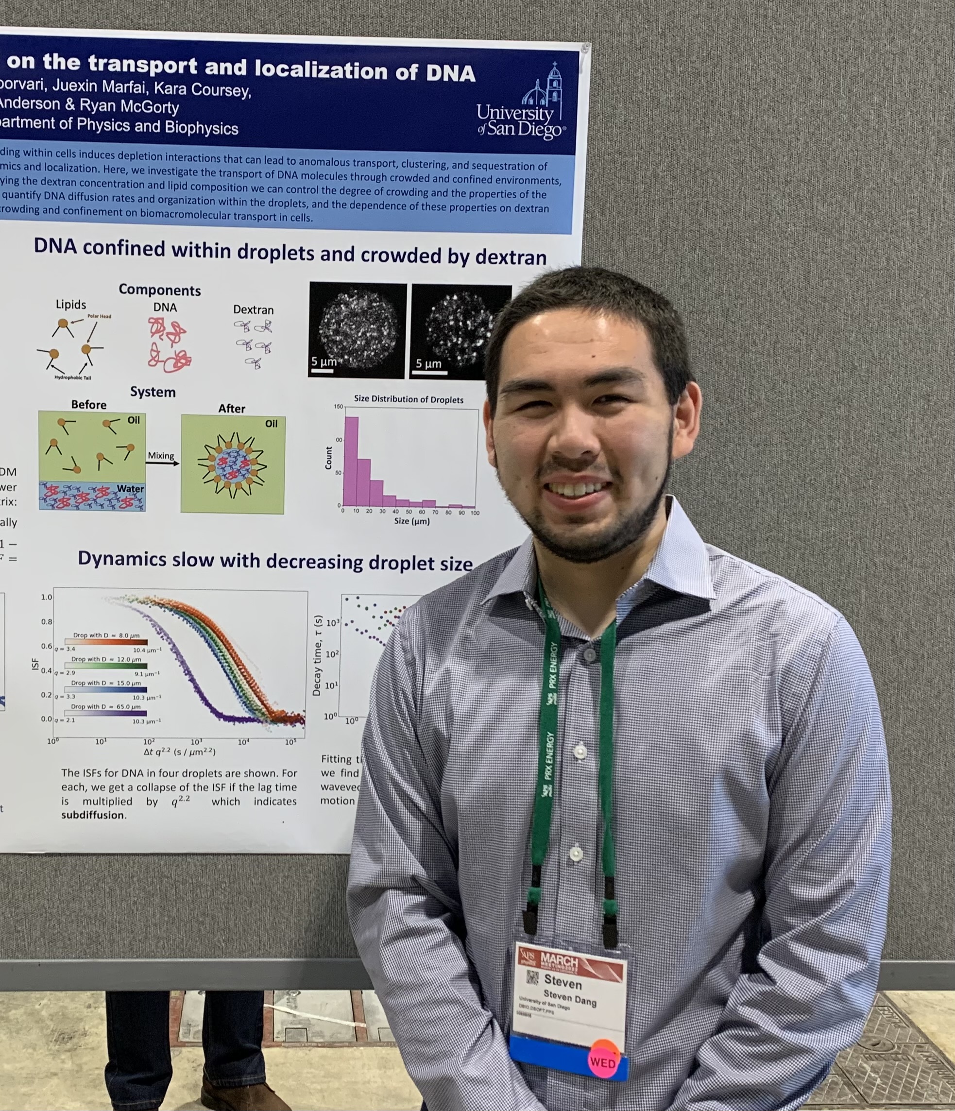
 | Crowding and confinement act in concert to slow DNA diffusion within cell-sized droplets *Mehdi Shafiei Aporvari*, *Steven Dang*, *Juexin Marfai*, Kara Coursey, *Ryan McGorty*, Rae M. Robertson-Anderson Published in iScience, September 2022 (see publication) Research themes: Differential Dynamic Microscopy |
 | Optical-Tweezers-integrating-Differential-Dynamic-Microscopy maps the spatiotemporal propagation of nonlinear strains in polymer blends and composites *Karthik R. Peddireddy*, Ryan Clairmont, *Philip Neill*, *Ryan McGorty*, Rae M. Robertson-Anderson Published in Nature Communications, September 2022 (see publication) Research themes: Differential Dynamic Microscopy |
 | Quantifying Cytoskeleton Dynamics Using Differential Dynamic Microscopy Hannah N. Verwei, Gloria Lee, *Gregor Leech*, Irene Isturiz Petitjean, Gijsje H. Koenderink, Rae M. Robertson-Anderson, *Ryan McGorty* Published in JoVE, June 2022 (see publication) Research themes: Cytoskeletal Networks, Differential Dynamic Microscopy |
| | Subtle changes in crosslinking drive diverse anomalous transport characteristics in actin-microtubule networks Sylas Anderson, *Jonathan Garamella*, Serenity Adalbert, *Ryan McGorty*, Rae M Robertson-Anderson Published in Soft Matter, March 2021 (see publication) Research themes: Cytoskeletal Networks, Differential Dynamic Microscopy |
 | Myosin-driven actin-microtubule networks exhibit self-organized contractile dynamics Gloria Lee, *Gregor Leech*, Michael J. Rust, Moumita Das, *Ryan McGorty*, Jennifer L. Ross, Rae M Robertson-Anderson Published in Science Advances, February 2021 (see publication) Research themes: Cytoskeletal Networks, Differential Dynamic Microscopy |
 | Two-color differential dynamic microscopy for capturing fast dynamics *Ruilin You*, *Ryan McGorty* Published in Review of Scientific Instruments, February 2021 (see publication) Research themes: Differential Dynamic Microscopy |
| | Topology-dependent anomalous dynamics of ring and linear DNA are sensitive to cytoskeleton crosslinking *Devynn Wulstein*, Kathryn Regan, *Jonathan Garamella*, *Ryan McGorty*, Rae M Robertson-Anderson Published in Science Advances, December 2019 (see publication) Research themes: Cytoskeletal Networks, Differential Dynamic Microscopy, Light Sheet Microscopy |
 | Filament Rigidity Vies with Mesh Size in Determining Anomalous Diffusion in Cytoskeleton Sylas J. Anderson, *Christelle Matsuda*, *Jonathan Garamella*, *Karthik Reddy Peddireddy*, Rae M Robertson-Anderson, *Ryan McGorty* Published in Biomacromolecules, November 2019 (see publication) Research themes: Cytoskeletal Networks, Differential Dynamic Microscopy, Light Sheet Microscopy |
| | Measuring capillary wave dynamics using differential dynamic microscopy *Jing Wang*, *Ryan McGorty* Published in Soft Matter, October 2019 (see publication) Research themes: Colloidal Fluids, Differential Dynamic Microscopy |
| | Bridging the spatiotemporal scales of macromolecular transport in crowded biomimetic systems Kathryn Regan, *Devynn Wulstein*, *Hannah Rasmussen*, *Ryan McGorty*, Rae M. Robertson-Anderson Published in Soft Matter, February 2019 (see publication) Research themes: Cytoskeletal Networks, Differential Dynamic Microscopy, Light Sheet Microscopy |
 | Point-spread function engineering enhances digital Fourier microscopy *Devynn Wulstein*, *Ryan McGorty* Published in Optics Letters, October 2017 (see publication) Research themes: Differential Dynamic Microscopy |
 | Light-sheet microscopy with digital Fourier analysis measures transport properties over large field-of-view *Devynn Wulstein*, Kathryn Regan, Rae M. Robertson-Anderson, *Ryan McGorty* Published in Optics Express, September 2016 (see publication) Research themes: Differential Dynamic Microscopy, Light Sheet Microscopy |
 | Dynamics and rheology at micro and macro scales of colloidal rod gels *Laura Morocho*, *Ryle Rel*, *Nikhil Sonthalia*, *Ryan McGorty* Presented at APS March Meeting, March 2025 (see published abstract) Research themes: Differential Dynamic Microscopy, Rheology |
| | Quantifying dynamics in 3D with light field and differential dynamic microscopy *Dylan Gage*, *Gildardo Martinez*, *Zayda Kellogg*, *Ryan McGorty* Presented at APS March Meeting, March 2025 (see published abstract) Research themes: Differential Dynamic Microscopy |
| | Characterizing diffusion in complex environments created by optical force fields using particle tracking and differential dynamic microscopy *Elijah Avery*, *Anindya Chowdhury*, *Ryan McGorty* Presented at APS March Meeting, March 2025 (see published abstract) Research themes: Differential Dynamic Microscopy |
| | Spatiotemporal mapping of non-equilibrium dynamics and mechanics in active cytoskeleton composites Aysan Razzaghi, *Elijah Avery*, Megan Valentine, *Ryan McGorty*, Rae Robertson-Anderson Presented at APS March Meeting, March 2025 (see published abstract) Research themes: Differential Dynamic Microscopy, Rheology |
 | Optical tweezers map spatiotemporal force generation in active actin-microtubule composites *Anindya Chowdhury*, *Karthik Reddy Peddireddy*, Moumita Das, Mehrzad Sasanpour, Jennifer Ross, *Ryan McGorty*, Megan T. Valentine, Michael J. Rust, Rae M. Robertson-Anderson Presented at APS March Meeting, March 2024 (see published abstract) Research themes: Differential Dynamic Microscopy |
| | Improved quantification of colloidal dynamics with machine learning and simulations *Dylan Gage*, *Gildardo Martinez*, *Justin Siu*, *Emma Kao*, *Juan Carlos Avila*, *Ruilin You*, *Ryan McGorty* Presented at APS March Meeting, March 2024 (see published abstract) Research themes: Differential Dynamic Microscopy |
| | Dynamics and rheology of active biomaterials and gels quantified with optical microscopy tools *Ryan McGorty* Presented at APS March Meeting, March 2024 (see published abstract) Research themes: Differential Dynamic Microscopy |
| | Dynamics and rheology of colloidal rod suspensions measured with differential dynamic microscopy *Laura G Morocho*, *Nikhil Sonthalia*, *Ryle R. Rel*, Esther Yang, *Ryan McGorty* Presented at APS March Meeting, March 2024 (see published abstract) Research themes: Differential Dynamic Microscopy, Rheology |
| | Quantifying dynamics of soft and active matter with microscopy and machine learning *Gildardo Martinez*, *Justin Siu*, *Dylan Gage*, *Emma Kao*, *Juan Carlos Avila*, *Ruilin You*, *Ryan McGorty* Presented at APS March Meeting, March 2024 (see published abstract) Research themes: Differential Dynamic Microscopy |
 | Nonlinear local straining leads to non-equilibrium deformation fields and dynamics of motor-driven cytoskeleton composites Mehrzad Sasanpour, Daisy H. Achiriloaie, Maya Hendija, *Karthik R. Peddireddy*, *Ryan McGorty*, Rae Robertson-Anderson Presented at APS March Meeting, March 2023 (see published abstract) Research themes: Cytoskeletal Networks, Differential Dynamic Microscopy |
| | Differential dynamic microscopy enhanced with convolutional neural networks *Justin Siu*, *Ruilin You*, *Steven Dang*, *Ryan McGorty* Presented at APS March Meeting, March 2023 (see published abstract) Research themes: Differential Dynamic Microscopy |
| | Nonlinear Mechanics and Stress Propagation in Out-of-Equilibrium Cytoskeleton Composites Daisy H. Achiriloaie, Kit K. Bennett, Mehrzad Sasanpour, *Karthik Peddireddy*, Michael J. Rust, Moumita Das, Megan T. Valentine, Janet Y. Sheung, Jennifer L. Ross, *Ryan McGorty*, Rae Robertson-Anderson Presented at APS March Meeting, March 2023 (see published abstract) Research themes: Cytoskeletal Networks, Differential Dynamic Microscopy |
| | Motor-driven advection competes with crowding to drive spatiotemporally heterogeneous transport in cytoskeleton composites Brian Y. Lee, Janet Y. Sheung, *Jonathan Garamella*, Stella Kahl, *Ryan McGorty*, Rae Robertson-Anderson Presented at APS March Meeting, March 2023 (see published abstract) Research themes: Cytoskeletal Networks, Differential Dynamic Microscopy |
| | Motor-driven advection competes with macromolecular crowding to drive spatiotemporally heterogeneous transport in cytoskeleton composites Janet Y. Sheung, *Jonathan Garamella*, Stella Kahl, Brian Y. Lee, Nadia Schwartz Bolef, Daisy H. Achiriloaie, Jennifer L. Ross, *Ryan McGorty*, Rae Robertson-Anderson Presented at APS March Meeting, March 2023 (see published abstract) Research themes: Cytoskeletal Networks, Differential Dynamic Microscopy |
| | DNA transport within confined motor-driven cytoskeletal networks *Mehdi Shafiei Aporvari*, *Philip Neill*, Daisy H. Achiriloaie, *Ryan McGorty*, Rae Robertson-Anderson Presented at APS March Meeting, March 2023 (see published abstract) Research themes: Cytoskeletal Networks, Differential Dynamic Microscopy |
| | Time-varying stress response and deformation dynamics of topologically-active DNA solutions *Karthik Peddireddy*, *Philip Neill*, *Juexin Marfai*, *Ryan McGorty*, Rae Robertson-Anderson Presented at APS March Meeting, March 2023 (see published abstract) Research themes: Differential Dynamic Microscopy |
 | Mapping nonlinear stress propagation in non-equilibrium complex fluids *Karthik Peddireddy*, Ryan Clairmont, *Philip Neill*, *Jonathan Garamella*, *Ryan McGorty*, Rae M. Robertson-Anderson Presented at APS March Meeting, March 2022 (see published abstract) Research themes: Differential Dynamic Microscopy |
| | DNA dynamics in crowded cell-sized droplets *Mehdi Shafiei Aporvari*, *Steven Dang*, *Juexin Marfai*, Kara Coursey, *Ryan McGorty*, Rae M. Robertson-Anderson Presented at APS March Meeting, March 2022 (see published abstract) Research themes: Cytoskeletal Networks, Differential Dynamic Microscopy |
| | Effects of crowding and confinement on the transport and localization of DNA *Steven Dang*, *Mehdi Shafiei Aporvari*, *Juexin Marfai*, Kara Coursey, *Ryan McGorty*, Rae M. Robertson-Anderson Presented at APS March Meeting, March 2022 (see published abstract) Research themes: Differential Dynamic Microscopy |
| | Tunable Non-Equilibrium Dynamics of Motor-Driven Cytoskeleton Composites Daisy H. Achiriloaie, Christopher Currie, Maya Hendija, *Ryan McGorty*, Jennifer L. Ross, Moumita Das, Michael J. Rust, Janet Y. Sheung, Rae M. Robertson-Anderson Presented at APS March Meeting, March 2022 (see published abstract) Research themes: Cytoskeletal Networks, Differential Dynamic Microscopy |
 | DNA transport and conformational dynamics in active cytoskeleton composites *Jonathan Garamella*, Serenity Adalbert, *Gina Aguirre*, *Ryan McGorty*, Rae M. Robertson-Anderson Presented at APS March Meeting, March 2021 (see published abstract) Research themes: Cytoskeletal Networks, Differential Dynamic Microscopy |
| | Mapping the nonlinear stress propagation in topological polymer blends *Karthik Peddireddy*, *Gina Aguirre*, *Jonathan Garamella*, *Ryan McGorty*, Rae M. Robertson-Anderson Presented at APS March Meeting, March 2021 (see published abstract) Research themes: Differential Dynamic Microscopy |
| | Differential Dynamic Microscopy of Active Actin-Microtubule Networks *Gregor Leech*, Gloria Lee, Michael Rust, Moumita Das, Jennifer L. Ross, *Ryan McGorty*, Rae M. Robertson-Anderson Presented at APS March Meeting, March 2021 (see published abstract) Research themes: Cytoskeletal Networks, Differential Dynamic Microscopy |
| | Tuning Dynamics of Myosin-Driven Actin-Microtubule Networks Gloria Lee, *Gregor Leech*, Christopher Currie, Michael Rust, Jennifer L. Ross, *Ryan McGorty*, Rae M. Robertson-Anderson Presented at APS March Meeting, March 2021 (see published abstract) Research themes: Cytoskeletal Networks, Differential Dynamic Microscopy |
 | Anomalous transport across scales in crosslinked actin-microtubule composites Sylas Anderson, *Jonathan Garamella*, *Ryan McGorty*, Rae M. Robertson-Anderson Presented at APS March Meeting, March 2020 (see published abstract) Research themes: Cytoskeletal Networks, Differential Dynamic Microscopy |
| | Non-ergodic transport and conformational dynamics of DNA in biomimetic cytoskeleton networks *Jonathan Garamella*, *Gina Aguirre*, Kathryn Regan, *Ryan McGorty*, Rae M. Robertson-Anderson Presented at APS March Meeting, March 2020 (see published abstract) Research themes: Cytoskeletal Networks, Differential Dynamic Microscopy |
| | Temporal super-resolution differential dynamics microscopy for detecting fast dynamics *Ruilin You*, *Ryan McGorty* Presented at APS March Meeting, March 2020 (see published abstract) Research themes: Differential Dynamic Microscopy |
| | Observing phase separation in colloid-polymer mixtures with a custom light-sheet rheoscope *Jing Wang*, *Ryan McGorty* Presented at APS March Meeting, March 2020 (see published abstract) Research themes: Colloidal Fluids, Differential Dynamic Microscopy |
| | Connecting microscale stresses to macromolecular motion in entangled ring-linear DNA blends *Karthik Peddireddy*, Megan C. Lee, *Jonathan Garamella*, *Ryan McGorty*, Rae M. Robertson-Anderson Presented at APS March Meeting, March 2020 (see published abstract) Research themes: Cytoskeletal Networks, Differential Dynamic Microscopy |
| Non-Ergodic Transport and Conformational Dynamics of DNA in Biomimetic Cytoskeleton Networks *Jonathan Garamella*, *Gina Aguirre*, *Ryan McGorty*, Rae M. Robertson-Anderson Presented at Biophysical Society Annual Meeting, February 2020 (see published abstract) Research themes: Cytoskeletal Networks, Differential Dynamic Microscopy |
 | Ensemble dynamics of large DNA molecules within entangled and crosslinked cytoskeleton networks *Devynn Wulstein*, Kathryn Regan, Shea Ricketts, Rae M. Robertson-Anderson, *Ryan McGorty* Presented at APS March Meeting, March 2019 (see published abstract) Research themes: Cytoskeletal Networks, Differential Dynamic Microscopy |
| | Measuring the dispersion relation of capillary waves using differential dynamic microscopy *Jing Wang*, *Ryan McGorty* Presented at APS March Meeting, March 2019 (see published abstract) Research themes: Colloidal Fluids, Differential Dynamic Microscopy |
| | Measure particle diffusion in cytoskeletal networks with light-sheet and differential dynamic microscopy *Christelle Matsuda*, Sylas Anderson, Rae M. Robertson-Anderson, *Ryan McGorty* Presented at APS March Meeting, March 2019 (see published abstract) Research themes: Cytoskeletal Networks, Differential Dynamic Microscopy |
| | Diffusion and Conformational Dynamics of Linear and Circular DNA in Crosslinked Cytoskeleton Composites Kathryn Regan, *Devynn Wulstein*, Shea Ricketts, *Ryan McGorty*, Rae M. Robertson-Anderson Presented at APS March Meeting, March 2019 (see published abstract) Research themes: Cytoskeletal Networks, Differential Dynamic Microscopy |
 | Selective-plane illumination microscopy to characterize diffusion of DNA in cytoskeletal networks *Devynn Wulstein*, Kathryn Regan, Shea Ricketts, Rae M. Robertson-Anderson, *Ryan McGorty* Presented at APS March Meeting, March 2018 (see published abstract) Research themes: Cytoskeletal Networks, Differential Dynamic Microscopy, Light Sheet Microscopy |
Published:
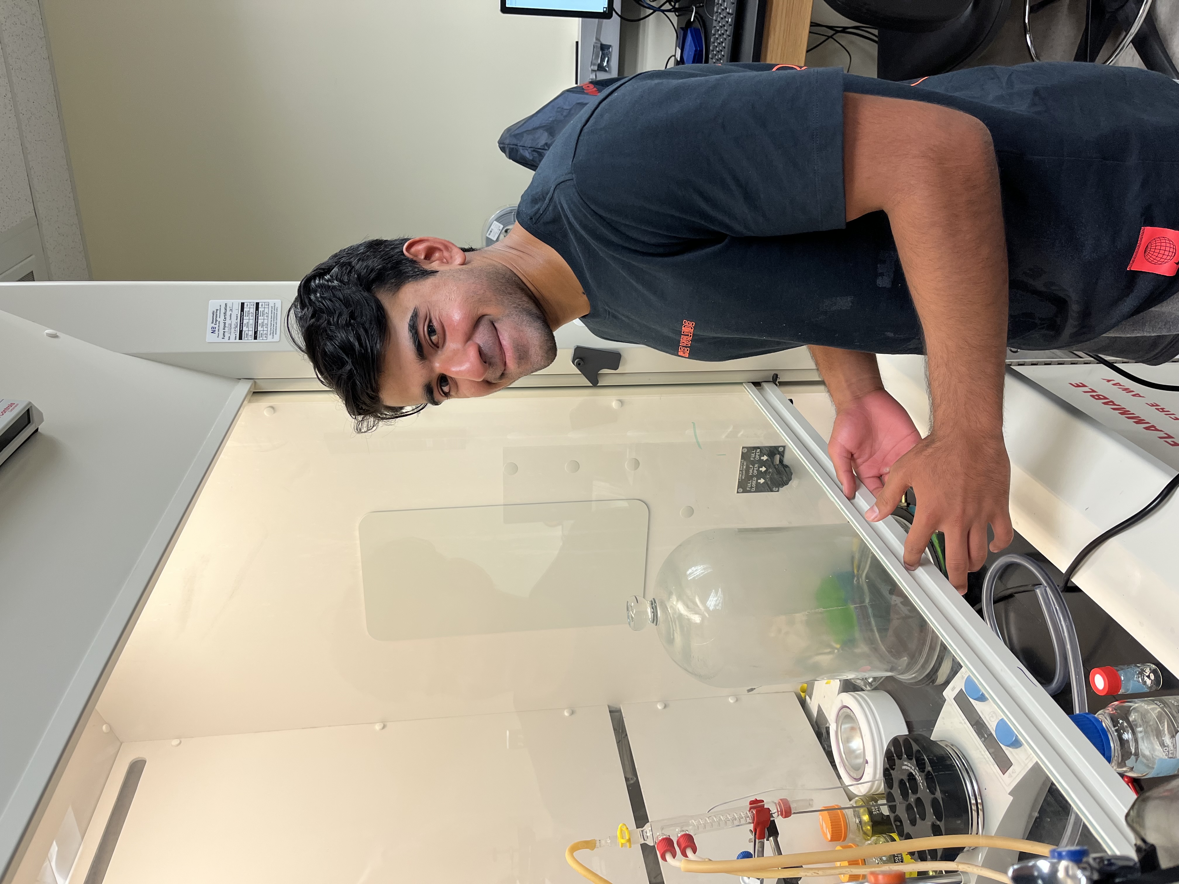
We have two light sheet microscopes in our lab.
Published:
Thanks to an NSF-MRI grant we have a DHR-3 rheometer which we use to study the rheology (ie, how materials flow or deform when a force is applied) of colloidal fluids, suspensions of DNA, and other materials.
Published:
| | Before the breach: Interactions between colloidal particles and liquid interfaces at nanoscale separations Anna Wang, Jos W. Zwanikken, David M. Kaz, *Ryan McGorty*, Aaron M. Goldfain, W. Benjamin Rogers, Vinothan N. Manoharan Published in Physical Review E, October 2019 (see publication) Research themes: Colloidal Fluids, Holographic Microscopy |
| | Contact-line pinning controls how quickly colloidal particles equilibrate with liquid interfaces Anna Wang, *Ryan McGorty*, David M. Kaz, Vinothan N. Manoharan Published in Soft Matter, November 2016 (see publication) Research themes: Colloidal Fluids, Holographic Microscopy |
Published:
Entangled polymers have complex viscoelastic properties that are tuned by polymer lengths and flexibilities. Entangled composites of distinct polymers offer added versatility and display nonlinear mechanics, serving as a platform for multifunctional materials. To determine the role of flexibility and length in polymer composites we use optical tweezers and confocal microscopy to measure mechanical and structural properties of co-entangled actin and DNA. Actin filaments have lengths of 5-20 μm, comparable to their persistence length, while DNA of similar lengths have hundreds of persistence lengths per chain. To characterize the nonlinear mechanics of actin-DNA composites, we optically drive a microsphere through the composite and measure the induced force during and following strain. We characterize viscoelasticity and relaxation timescales; and determine the dependence of these quantities on the actin:DNA ratio (0:1-1:0) and DNA length (4-100 μm). We use confocal microscopy to image distinctly-labeled co-entangled actin and DNA and characterize network homogeneity and fluctuations. Initial results show actin and DNA are well-integrated and form structurally homogenous networks that exhibit stiffness and relaxation times that increase nonlinearly with increased actin.
Download here
Published:
The high concentrations of proteins crowding cells greatly influence intracellular DNA dynamics. These crowders, ranging from small mobile proteins to large cytoskeletal filaments such as semiflexible actin and rigid microtubules, can hinder diffusion and induce conformational changes in DNA. The rigidity, mobility, and concentration of crowders all play a role in DNA transport, yet previous studies have mainly focused on the effect of small mobile crowders on transport. At the same time the rigid cytoskeleton has been identified as a key factor suppressing viral transfection and gene delivery. Here, we use fluorescence microscopy and custom single-molecule conformational tracking algorithms to measure center-of-mass transport and time-varying conformational sizes and shapes of single 115 kbp DNA molecules diffusing in networks of actin filaments and microtubules. We determine the dependence of protein concentration (6 – 23 μM) and rigidity (actin vs microtubules) on DNA dynamics. Corresponding measurements with monomeric actin and tubulin identify the roles that network rigidity versus excluded volume play in transport. Initial results show that crowding by microtubules induces anomalous transport and larger, slower conformational fluctuations of DNA.
Download here
Published:
Fiber networks are present in various systems from textiles and paper to the cytoskeleton and tissues. Simulations of fiber networks have unveiled interesting emergent phenomena. However, it has proven difficult to experimentally probe how forces are distributed across individual fibers in such networks. Here we are able to determine the force distribution through multiple geometries of macroscopic networks in both two and three dimensions in a simple manner. We construct networks out of macroscopic strips of conductive fabric. We measure the change in resistivity of each element of conductive fabric as a network is perturbed to determine the force on each element simultaneously. We investigate the magnitudes of the forces within the network and how those forces are spatially distributed through the network. Our model system makes determining the stress throughout a large system quite simple, and can be applied to large and three-dimensional systems.
Download here
Published:
How polymers such as DNA move through crowded cytoskeletal environments has yet to be fully understood. New techniques to quantitatively characterize how the dynamics may differ from simple diffusion and to link those anomalous dynamics of DNA to properties of the crowding cytoskeletal network are required. Here we demonstrate a technique that measures the ensemble and single molecule dynamics over a large range of time and length scales. We use selective-plane illumination microscopy (SPIM) to observe the dynamics of fluorescently labeled DNA molecules in varying networks of actin and microtubules. Due to the Gaussian nature of the excitation light-sheet, we use single molecule tracking in the region with high optical sectioning and capture ensemble dynamics in the regions with low optical sectioning. We use differential dynamic microscopy (DDM) to analyze ensemble dynamics. Using SPIM and DDM, we efficiently obtain single-molecule and ensemble dynamics from the same time-series of images. Additionally, we can image and measure the dynamics of the three-dimensional network. Our use of single-molecule tracking and DDM on the same image acquired with SPIM could be extended to characterizing in vivo dynamics or other complex fluids with non-ergodic behavior.
Download here
Published:
We present a highly-flexible, cost-effective multimodal microscope using a programmable electric paper display. We demonstrate its ability to acquire dark-field and phase contrast images. The display can be used with either incoherent or laser illumination.
Download here
Published:
In order to carry out key cellular processes, DNA molecules must diffuse through the crowded cytoskeletal network, comprised of thin semiflexible actin filaments and thick rigid microtubules. Each of these cytoskeletal filaments can also be crosslinked to varying degrees, altering the network structure and dynamics and hence the impact on DNA diffusion and conformational dynamics. Here, we use single-molecule conformational tracking to investigate the effect of cytoskeleton crosslinking on the transport properties and time-varying conformations of diffusing DNA molecules. Specifically, we track the center-of-mass, size, and shape of single linear and circular DNA molecules diffusing in crosslinked composites of actin and microtubules. We quantify transport coefficients, anomalous diffusion scaling exponents, lengths and timescales of intramolecular fluctuations, and shape and size dynamics of DNA. We determine the role of DNA topology (circular vs linear) as well as cytoskeleton crosslinking motif on DNA dynamics and conformations. Our results shed light on how macromolecular topology and network structure impact macromolecular mobility in crowded cell-like environments.
Download here
Published:
A paper-based microscope makes for a rugged and versatile solution to bringing science and science education to a variety of settings including those that are resource-scarce. This has been well demonstrated by the Foldscope project. Here, we describe an extension to that idea. We designed a paper-based method to control the sample illumination in a portable microscope. Controlling the illumination of the sample allows us to employ a variety of microscope imaging modalities including bright-field, dark-field and phase contrast. With this instrument, students can explore those different techniques. Additionally, it can be used to perform advanced imaging, including quantitative phase imaging, in the field, away from advanced and costly instrumentation.
Download here
Published:
Filamentous actin and microtubules contribute to the crowded environment of the cytoplasm. Modeling the cytoplasm, we create networks of varying crosslinking motifs and varying ratios of actin and microtubules. We crosslink either actin to actin, tubulin to tubulin, or tubulin to actin. We add micron-sized fluorescent beads to the various networks, record videos of the samples using light-sheet microscopy, and perform differential dynamic microscopy (DDM) analysis. We compare DDM and single-particle tracking results to examine differences between ensemble and single-particle transport properties. We find varying degrees of anomalous diffusion and significant heterogeneity in the dynamics, particularly in actin environments.
Download here
Published:
The diffusion of microscopic particles through the cell, important to processes such as transcription, viral infection, transfection and gene delivery, is largely controlled by the complex cytoskeletal network that pervades the cytoplasm. The cytoskeleton is predominantly made up of thin semiflexible actin filaments and thicker, more rigid microtubules, as well as binding proteins that can crosslink each filament. By varying the relative concentrations of actin and microtubules, as well as the degree to which each filament is crosslinked, the cytoskeleton can display a host of different structural and dynamic properties that in turn impact the diffusion of particles through the network. Here we use single particle tracking methods to quantify the mean-squared displacements of microspheres diffusing in custom-designed in vitro composites of actin and microtubules. We show that particles exhibit subdiffusion, with scaling exponents and transport coefficients that decrease as the relative fraction of actin in composites increases. By evaluating the distributions of bead displacements, we also find that composites induce unique non-gaussian diffusion characteristics and substantial heterogeneities in particle trajectories.
Download here
Published:
Through the use of a colloid-polymer system employing temperature-sensitive pNIPAM microgel colloidal particles, we observe the nucleation and dissolution of colloid-rich liquid droplets. We use light-sheet microscopy to observe the formation and dissolution of colloid-rich droplets in three-dimensions and with optical sectioning. Our colloid-polymer system allows us to precisely tune the equilibrium state–mixed or demixed–by adjusting the sample temperature. With videos obtained from the light-sheet microscope, we perform image analysis of fluctuating droplets to extract the surface tension.
Download here
Published:
With a recently-developed method for measuring capillary wave dynamics at the interface of fluids we examine non-equilibrium interfacial fluctuations. We use a colloidal-polymer mixture that separates into colloid-rich and colloid-poor fluid phases with an ultra-low surface tension and capillary velocities on the order of a micron per second. We use differential dynamic microscopy (DDM) to measure the two-dimensional dispersion relation of capillary waves at spatial frequencies spanning over an order of magnitude. Using temperature-responsive colloidal particles (pNIPAM) to tune the phase diagram we investigate the interfacial fluctuations between non-equilibrium phases.
Download here
Published:
The cellular interior is highly crowded. How biological macromolecules, such as DNA, diffuse through these environments has yet to be fully elucidated. We mimic the crowded cellular environment by creating custom-designed co-polymerized networks of actin and microtubules that are crosslinked at various motifs. We study the effect of the co-entangled and co-crosslinked cytoskeleton networks on the ensemble dynamics of large circular and linear DNA molecules using selective-plane illumination differential dynamic microscopy (SPIDDM). As a digital Fourier microscopy technique, SPIDDM measures dynamics over a large range of length and time scales that supplement and expand on the dynamics measured using single-molecule tracking of DNA in the same environments. We find interesting differences between ensemble and single-molecule dynamics over the temporal and spatial scales probed.
Download here
Published:
Macromolecular crowding is an increasingly relevant biophysical phenomenon affecting gene expression, drug delivery, protein function, and intracellular transport. Of particular interest is the influence that cytoskeletal filaments have on the transport and conformational dynamics of large DNA molecules–two properties critical for processes such as transfection, viral infection, and gene therapy. Here, we use single-molecule conformational tracking (SMCT) to elucidate the transport properties and conformational dynamics of linear and relaxed circular (ring) DNA in in vitro composite networks of actin and microtubules with variable types of crosslinking. Specifically, we investigate the impact of crosslinking actin to actin, microtubules to microtubules, and actin to microtubules. While both linear and ring DNA undergo anomalous subdiffusion in all networks, the transport properties are heavily influenced by DNA topology (linear vs ring).
Download here
Published:
Ring polymers, as well as mixtures of ring and linear polymers, are ubiquitous in nature yet still poorly understood. Due to the lack of free ends in ring polymers, the motion and dynamics of entangled rings is complex and distinctly different than their linear chain counterparts. As such, the dynamics of entangled blends of ring and linear polymers remain a topic of fervent debate. Here, we use DNA - which occurs naturally in rings and linear forms - as a model system to investigate highly entangled ring-linear blends. To elucidate the dynamics of these blends, we demonstrate a novel technique that combines optical tweezers microrheology with fluorescence imaging and differential dynamic microscopy. This technique enables us to directly image single polymers while performing active microrheology. As a result, we show that it is possible to unambiguously connect the stresses induced by both linear and nonlinear strains to the corresponding macromolecular deformations and network rearrangement in ring-linear polymer blends.
Download here
Published:
We study liquid-liquid phase separation (LLPS) with a colloid-polymer system subjected to shear. Our colloid-polymer mixture consists of temperature-responsive PNIPAM microgel particles and polymers acting as a depletant. This mixture separates into two phases: a colloid-poor, or “gas” phase, and a colloid-rich, or “liquid” phase. We observe the process of phase separation using a custom-built light-sheet microscope, which allows for simultaneously acquiring optically-sectioned images of our sample and shearing the sample in a Couette geometry. We measure the size and shape of elongated liquid domains that have been deformed due to flow as a function of shear rate. The temperature-responsive feature of our colloidal particles allows us to further explore the kinetics of phase separation under shear flow. We hope our study of phase separation under shear can provide fundamental insights into hydrodynamics and thermodynamics and provide novel strategies for structuring soft materials.
Download here
Published:
We introduce dual-color differential dynamic microscopy (DDM) for detecting fast dynamics. DDM has been used extensively to measure the diffusive or ballistic motion of small particles, macromolecules and bacteria. Rather than localizing and tracking individual particles, DDM works by measuring the intensity fluctuations in images across a range of detectable spatial frequencies and provides data similar to that provided by dynamic light scattering. However, DDM is limited by the camera frame rate. Fast dynamics can be measured with high-speed cameras but those are typically expensive. We have developed a dual-color imaging setup which allows us to detect dynamics faster than the camera’s frame rate. We trigger blue and red light at well-defined times within a single image exposure. By analyzing each color channel separately and in combination we detect dynamics that are several times faster than the camera frame rate.
Download here
Published:
Macromolecular crowding is an increasingly relevant biophysical phenomenon that affects drug delivery, protein function, and intracellular transport. Of particular interest is the influence that cytoskeletal filaments have on the transport and conformational dynamics of large DNA molecules. Here, we use single-molecule conformational tracking (SMCT) to elucidate the transport properties and conformational dynamics of linear and relaxed circular (ring) DNA in in vitro composite networks of actin and microtubules with variable types of crosslinking. Specifically, we investigate the impact of crosslinking actin to actin, microtubules to microtubules, and actin to microtubules. While both linear and ring DNA undergo anomalous subdiffusion in all networks, the transport properties are heavily influenced by DNA topology. Linear DNA chains are compacted and display a single mode of subdiffusion, while ring DNA polymers are swollen and exhibit biphasic subdiffusion suggestive of transient threading by the biomimetic cytoskeleton. These results are bolstered by non-Gaussian van Hove distributions and non-ergodic behavior of both DNAs, with the transport of ring DNA molecules becoming less ergodic than their linear counterparts at longer times.
Download here
Published:
We use colloidal microgel particles, PNIPAM and PNIPMAM, in a colloid-polymer system to study phase separation. We synthesize particles of various sizes and characterize their temperature-tunable size with differential dynamic microscopy. When these colloidal particles are mixed with a depletant we observe fluid-fluid phase separation as well as gelation. The process of phase separation is observed using a custom-built light-sheet microscope. With a two-color fluorescent labeling scheme we can track single colloidal particles as well as the phases that emerge as an originally-mixed sample phase separates. Furthermore, we observe our samples under shear in both a custom-built cylindrical shear cell with our light-sheet microscope and in a commercial rheometer using brightfield microscopy. Studying the behavior of multiphase systems like our colloid-polymer samples strengthens our understanding of non-equilibrium thermodynamics while providing novel routes for structuring complex fluids.
Download here
Published:
The diffusion of microscopic particles through the cell is largely controlled by the cytoskeletal network, comprised of semiflexible actin filaments, rigid microtubules, and crosslinking proteins. Yet, how the interactions between actin and microtubules and the various types of filament crosslinking affect particle transport remain unresolved. In our experiments, we couple single-particle tracking (SPT) with differential dynamic microscopy (DDM) to characterize the transport of micron-sized particles diffusing through crosslinked composite networks of actin filaments and microtubules. Specifically, we investigate the impact of permanently crosslinking actin to actin, microtubules to microtubules, and actin to microtubules. By combining SPT and DDM, we are able to couple single-particle dynamics to ensemble transport phenomena, and link particle diffusion to the viscoelastic characteristics of the networks. We find that subtle changes to the crosslinking interactions between cytoskeleton filaments play surprisingly important roles in the anomalous subdiffusion that particles exhibit within a composite cytoskeletal system.
Download here
Published:
In this work, we explore the role of shear in structuring colloidal suspensions and how such shear-induced structuring affects rheological properties. We study mixtures of temperature-responsive poly-N-isopropylacrylamide (PNIPAm) particles about 600 nm in diameter and large polymer molecules acting as depletants. These mixtures undergo fluid-fluid phase separation or gelation depending on colloid and polymer concentrations.
Published:
The cytoskeleton, composed of actin, microtubules, and associated motor and binding proteins, is an actively rearranging network with tunable structure and mechanics. For example, myosin motors can induce contraction, extension and flow of actin networks. While actomyosin networks have been well-characterized, the impact of microtubules on actomyosin dynamics remains poorly understood. Here, we create active composite networks of actin and microtubules that exhibit contractile dynamics driven by myosin II. We tune the dynamics, activity and structure of composites by varying the relative concentrations of actin, microtubules, and myosin. Using multi-spectral confocal microscopy, along with differential dynamic microscopy (DDM), particle image velocimetry (PIV), and spatial image autocorrelation (SIA) analyses, we characterize how network composition affects the non-equilibrium dynamics and structure over time. Specifically, we use DDM and PIV to quantify the rate and directionality of network motion, and we use SIA to quantify time-varying network correlation length scales. These results provide new insight into the diverse interactions cells can use to tune activity, as well as the design of tunable active materials.
Download here
Published:
The cytoskeleton is a dynamic network of protein filaments and motors, including actin, microtubules and myosin, that enable key processes in the cell such as growth, movement, and cell division. Active rearrangement and contraction of actin networks by myosin motors has been extensively studied in recent years. However, how the interactions between actin and microtubules affects actomyosin activity remains poorly understood. We investigate the active dynamics and restructuring of composite networks of actin and microtubules driven by myosin II motors. We combine spatial image autocorrelation analysis and particle image velocimetry to characterize the time-varying network structure and velocity profiles of actin and microtubules. We show that actin and microtubules exhibit similar contractile flow fields that increase in magnitude and heterogeneity as the concentration of actin or myosin increases. We also find that the correlation length scales of the composite networks generally decrease over time but at a slower rate with higher concentrations of actin or myosin. These findings provide insight into how the cytoskeleton can tune its dynamics and structure to impact cellular processes such as movement and growth.
Download here
Published:
To connect the structural and flow properties of colloidal gels we experimentally probe colloid-polymer mixtures on a rheometer with an optical microscopy attachment. We study mixtures of a polymer and temperature-responsive hydrogel particles (pNIPAM) which undergo fluid-fluid phase separation or gelation at room temperature depending on concentrations. At higher temperatures (above about 32 °C) the hydrogel particles form a gel. We observe these mixtures as they are sheared in a parallel-plate geometry with an optically transparent base plate and a small gap thickness. At room temperature, we observe flow-aligned domains with shear-rate-dependent dimensions. Above 32 °C, the stronger interparticle attraction leads to vorticity-aligned flocs. We can cycle between flow-aligned and vorticity-aligned domains by varying the temperature by just a couple degrees. This allows us to investigate the formation of these shear-induced domains and to connect their spatial characteristics with the bulk sample flow properties.
Download here
Published:
The cytoskeleton is a non-equilibrium network of protein filaments, including semiflexible actin and rigid microtubules, as well as associated motor proteins that can dynamically rearrange the filaments. Here, we create co-entangled networks of microtubules and actin that exhibit active contractile dynamics driven by myosin II motors. We use multi-spectral confocal fluorescence microscopy and differential dynamic microscopy to determine the time-varying fluctuation decay times and contraction rates of the networks. We vary the concentrations of actin, microtubules and myosin to tune the dynamics from slow, organized contraction to disordered vortical flow. We also examine the impact of sample age on the non-equilibrium dynamics. Our findings shed light onto how the active composite cytoskeleton can finely tune its dynamics by varying the interactions between its constituents.
Download here
Published:
Light sheet fluorescence microscopy (LSFM) is emerging as a popular volumetric imaging method to study living cells, tissues, and whole organisms. It minimizes potential photodamage to the sample by confining the excitation light to the focal plane. However, most implementations of LSFM require two objective lenses placed orthogonally to one another. Such a setup precludes the use of high numerical aperture lenses and of many common sample mounting methods, such as between a glass slide and coverslip. Here, we adopt a single objective LSFM design. A single objective is used to both illuminate and image our sample. Downstream remote focusing lenses allow us to image the plane coincident with the illumination plane. With this configuration we are able to use a high numerical aperture objective lens (NA > 1.0) and acquire volumetric imaging data at several Hz. We show that this configuration and other LSFM setups are well suited to imaging colloidal samples. Particularly, we show that LSFM can be used to image colloidal and other soft matter samples while those samples are mechanically perturbed.
Download here
Published:
Entangled polymers and blends of varying topologies and stiffnesses exhibit complex nonlinear rheological properties that depend on the blend composition and the scale of the strain. How local nonlinear stresses propagate through these systems remains an open question. Here, we combine optical tweezers microrheology with fluorescence imaging and differential dynamic microscopy (DDM) to map the deformation field arising from a local nonlinear strain in entangled polymer blends. We use optical tweezers to impart local nonlinear strains and measure the resulting stresses during and following strain. We simultaneously image labeled DNA molecules surrounding the strain site and use DDM to determine how the macromolecular dynamics vary with distance from the applied strain. We perform these measurements on entangled solutions of linear and ring DNA, as well as blends of DNA and microtubules. Our approach, which combines active microrheology with macromolecular tracking and differential dynamic microscopy, directly links nonlinear stress propagation to macromolecular deformations and dynamics, and is applicable to a wide range of complex fluids and polymeric materials.
Download here
Published:
The cytoskeleton plays a key role in governing the intracellular transport of macromolecules and, in turn, controlling biophysical phenomena such as drug delivery, protein function, and transfection. While the impact of crowding by cytoskeleton filaments, including actin and microtubules, has been widely studied, how the non-equilibrium dynamics of cytoskeletal networks driven by motor proteins impact intracellular transport is oft overlooked. Here, we couple single-molecule conformational tracking (SMCT) and differential dynamic microscopy (DDM) to elucidate the transport and conformational dynamics of ring and linear DNA molecules crowded by composite networks of actin and microtubules undergoing active myosin-driven rearrangement. Using DDM, we quantify the ensemble dynamics of the DNA, finding the transport properties (subdiffusive, ballistic, etc.) depend on both DNA size and topology. With SMCT, we measure dynamics that transition between subdiffusive and ballistic, and determine how ring and linear DNA molecules are conformationally affected by interactions with the active cytoskeleton networks.
Download here
Published:
Motor-driven scaffolds of actin filaments and microtubules display rich non-equilibrium dynamics and structure that are critical to mesoscale cellular behavior. Numerous studies have investigated the dynamics of actomyosin networks, active kinesin-microtubule systems, and steady-state actin-microtubule composites. Yet, in cells all of these components interact to enable the diverse processes and properties that cells exhibit. Here, we create in vitro actin-microtubule networks driven by myosin-II minifilaments and quad-headed kinesin clusters, and use multi-color confocal microscopy to image the active composites. To characterize the non-equilibrium dynamics of actin and microtubules in the composites, we use differential dynamic microscopy (DDM) to quantify filament contraction and flow as the composites dynamically restructure. We find that myosin and kinesin activity lead to dramatically different dynamics of both filaments, and that static crosslinking of either filament changes the patterns and rates of motor-driven restructuring.
Download here
Published:
Two hallmarks of the intracellular environment are crowding and confinement. Macromolecular crowding within cells induces depletion interactions that can lead to anomalous transport, clustering, and sequestration of macromolecules such as DNA. Confinement by the lipid membrane of cells further dictates macromolecular dynamics and localization. Here, we investigate the transport of DNA molecules through crowded and confined environments, using dextran polymers as crowders and using lipids to encapsulate DNA and dextran in cell-sized droplets. By varying the dextran concentration and lipid composition we can control the degree of crowding and the properties of the confining boundary. We use differential dynamic microscopy (DDM) and spatial image autocorrelation analysis to quantify DNA diffusion rates and organization within the droplets, and the dependence of these properties on dextran concentration as well as droplet size and stiffness. Our results shed light on the effects on the coupled effects of crowding and confinement on biomacromolecular transport in cells.
Download here
Published:
We measure capillary wave fluctuations, droplet coalescence, and dripping in a colloidal fluid system by using a custom-built single objective light sheet microscope (SOLS). We acquire volumetric imaging data at several volumes per second with submicron resolution. We show that we can use SOLS to detect capillary fluctuations at the interface of coexisting colloidal fluids. We measure submicron fluctuations and capillary velocities on the order of 0.1 micron per second. We additionally measure the width of the growing bridge that connects a coalescing droplet to the continuous phase. Our SOLS system allows us to acquire fast optically sectioned images for volumetric data collection without needing to move the objective lens. Rather, the focal plane and sheet of excitation light are swept across the sample using a scanning mirror. This method provides fast 3D imaging with minimal mechanical perturbation to the sample.
Download here
Published:
We investigate the rheology, structure, and flow-induced patterning of colloidal gels composed of thermosensitive poly(N-isopropylacrylamide) (pNIPAM) microgel particles. We mix these particles with a polymer to induce a depletion attraction. At room temperature, these mixtures phase separate into coexisting fluid phases. At slightly higher temperatures, these mixtures form a gel due to the increased hydrophobicity of the colloidal microgel particles. By applying shear to our samples while in the fluid-fluid demixed state, we can deform the colloid-rich domains. By then increasing the temperature, these colloid-rich domains connect together to form a sample-spanning gel. We observe the flow-induced patterns that form when we continue shearing this gel and it yields. We find that the patterns depend on how deformed, by flow, the colloid-rich domains were prior to gelation. For a range of shear rates applied to the sample while in the fluid-fluid demixed state, when the sample transitions to the gel state and is sheared we observe vorticity-aligned log-like flocs which roll between the parallel plates of our geometry. Our data suggests that the morphology of the colloidal clusters, which is dependent on the shear rate applied while in the fluid-fluid demixed state, is an important factor in determining whether or not vorticity-aligned flocs form. This work may offer routes for tuning the strength and structure of colloidal gels.
Download here
Published:
Polymer topology has been shown to play a principal role in the rheology and miscibility of polymer composites. At the same time, active materials, which undergo bulk rheological changes driven by macromolecular rearrangement and restructuring, are the topic of intense investigation. Here, we design polymer composites comprising entangled ring DNA and dextran polymers, and measure the dependence of rheological properties on the ratio of the two polymers. To push the composites out of equilibrium we incorporate enzymes that convert DNA rings to linear fragments and measure the time-dependent rheological properties of the topologically-active DNA-dextran composites during enzymatic activity. The bulk linear viscoelastic moduli that we measure show that composites undergo shear thickening and thinning over time, with the timescale and magnitude of the rheological changes dependent on the DNA:dextran ratio. Our system combines the tunability and versatility of polymer composites with the power of topologically-distinct DNA and enzymatic reactions to create topologically-active polymeric fluids that can be used for diverse applications from drug delivery, to filtration to infrastructure repair.
Download here
Published:
Understanding the transport and conformational dynamics of biological macromolecules such as DNA in crowded and confined environments is critical to understanding biological processes such as viral infection, liquid-liquid phase separation, and transcription. Here we investigate the dynamics, conformations, and organization of DNA molecules confined in cell-sized droplets and crowded by dextran polymers. Specifically, we use differential dynamic microscopy to measure ensemble DNA dynamics and single-molecule conformational tracking to measure DNA trajectories, shapes, and localization. We directly connect these measurements to the microrheological properties of the crowded, confined environment by performing similar measurements with microspheres. We determine the impact of droplet size and crowding conditions, as well as DNA length and topology, on the measured transport and structural properties.
Download here
Published:
Topologically-novel entangled polymers and enzyme-driven active matter systems are examples of complex fluids that exhibit intriguing spatiotemporally varying responses to strain. Yet, how local nonlinear stresses propagate through these soft systems remains an open question. Here, we combine optical tweezers microrheology (OTM) with differential dynamic microscopy (DDM) to map the time-dependent deformation fields arising from local nonlinear straining in ‘topologically-active’ DNA fluids. Specifically, we measure the stresses imposed in the fluids by local nonlinear strains and simultaneously image labeled DNA molecules surrounding the strain site. Using DDM, we characterize the macromolecular dynamics and deformation as a function of strain rate and distance from the applied strain. We perform these measurements on blends of circular DNA undergoing enzymatically-driven topological conversion and fragmentation. Our coupled OTM-DDM platform directly links nonlinear stress propagation to macromolecular dynamics and network structure and is applicable to a wide range of active and heterogeneous complex fluids.
Download here
Published:
Download here
Published:
Download here
Published:
Download here
Published:
Download here
Published:
Download here
Published:
Download here
Published:
Download here
Published:
Download here
Published:
Download here
Published:
Download here
Published:
Download here
Published:
Download here
Published:
Download here
Published:
Download here
Published:
Download here
Published:
Download here
Published:
Download here
Published:
Download here
Published:
Download here
Published:
Download here
Published:
Download here
Published:
Download here
Published:
Download here
Published:
Download here
Published:
Download here
Published:
Download here
Published:
Download here
Published:
Download here
Published:
Download here
Published:
Download here
Published:
Download here
Published:
Download here
Published:
Download here
Published:
Download here
Physics 136, 136L, & 481W, University of San Diego, 2015
…
Physics 136, 136L, 137, 270, & 270L, University of San Diego, 2016
…
Physics 381W, 270, & 494, University of San Diego, 2017
…
Physics 136, 270, 494, 381, University of San Diego, 2018
…
Physics 270, 270L, 281, 314, & 340, University of San Diego, 2019
…
Physics 270, 281, 381, & 494, University of San Diego, 2020
…
Physics 270 & 494, University of San Diego, 2022
…
Physics 381 & 340, University of San Diego, 2023
…
Physics 400 & 270, University of San Diego, 2024
…
Physics 381 & 340, University of San Diego, 2025
…
Published:
Ryan’s CV is available here (updated 11/2024).
Published:
Currently pursuing a PhD in marine biology at the University of Hawai'i.
Published:
Currently ...
Published:
Andrew is currently in pharmacy school at the University of the Pacific.
Published:
Currently ...
Published:
Currently ...
Published:
Published:
Currently pursuing a PhD in physics at the University of Nevada, Reno.
Published:
Currently pursuing a PhD in physics at Emory University.
Published:
Published:
Published:
Published:
Published:
Published:
Published:
Published:
Published:
Published:
Published:
Published:
Published:
Currently pursuing a PhD at the University of Arizona College of Optical Sciences.
Published:
Published:
Currently at LumiraDX.
Published:
Published:
Published:
Published:
Currently working as an analyst at California Business Advisors.
Published:
Published:
Published:
Published:
Published:
Published:
Published:
| | Protocol for analyzing DNA dynamics in the presence of crowders and confined within cell-sized droplets *Mehdi Aporvari*, *Ryan McGorty*, Rae M. Robertson-Anderson Published in STAR Protocols, September 2024 (see publication) Research themes: Differential Dynamic Microscopy |
| | Crowding and confinement act in concert to slow DNA diffusion within cell-sized droplets *Mehdi Shafiei Aporvari*, *Steven Dang*, *Juexin Marfai*, Kara Coursey, *Ryan McGorty*, Rae M. Robertson-Anderson Published in iScience, September 2022 (see publication) Research themes: Differential Dynamic Microscopy |
| | DNA transport within confined motor-driven cytoskeletal networks *Mehdi Shafiei Aporvari*, *Philip Neill*, Daisy H. Achiriloaie, *Ryan McGorty*, Rae Robertson-Anderson Presented at APS March Meeting, March 2023 (see published abstract) Research themes: Cytoskeletal Networks, Differential Dynamic Microscopy |
| | DNA dynamics in crowded cell-sized droplets *Mehdi Shafiei Aporvari*, *Steven Dang*, *Juexin Marfai*, Kara Coursey, *Ryan McGorty*, Rae M. Robertson-Anderson Presented at APS March Meeting, March 2022 (see published abstract) Research themes: Cytoskeletal Networks, Differential Dynamic Microscopy |
| | Effects of crowding and confinement on the transport and localization of DNA *Steven Dang*, *Mehdi Shafiei Aporvari*, *Juexin Marfai*, Kara Coursey, *Ryan McGorty*, Rae M. Robertson-Anderson Presented at APS March Meeting, March 2022 (see published abstract) Research themes: Differential Dynamic Microscopy |
Published:
Published:
Published:
Published:
Currently pursuing a PhD at UCSD
Published:
Published:
Published:
Published:
Published:
Published:
Published:
Published:
Published:
Published:
Published:
| | Characterizing diffusion in complex environments created by optical force fields using particle tracking and differential dynamic microscopy *Elijah Avery*, *Anindya Chowdhury*, *Ryan McGorty* Presented at APS March Meeting, March 2025 (see published abstract) Research themes: Differential Dynamic Microscopy |
| | Optical tweezers map spatiotemporal force generation in active actin-microtubule composites *Anindya Chowdhury*, *Karthik Reddy Peddireddy*, Moumita Das, Mehrzad Sasanpour, Jennifer Ross, *Ryan McGorty*, Megan T. Valentine, Michael J. Rust, Rae M. Robertson-Anderson Presented at APS March Meeting, March 2024 (see published abstract) Research themes: Differential Dynamic Microscopy |
Published:
Published:
Published:
Published:
Published:
Published:
Published:
Published:
Published:
Published: